Host-fungus interactions have gained a lot of interest in the past few decades, mainly due to an increasing number of fungal infections that are often associated with a high mortality rate in the absence of effective therapies. These interactions can be studied at the genetic level or at the functional level via imaging. Here, we introduce a new image processing method that quantifies the interaction between host cells and fungal invaders, for example, alveolar macrophages and the conidia of Aspergillus fumigatus. The new technique relies on the information content of transmitted light bright field microscopy images, utilizing the Hessian matrix eigenvalues to distinguish between unstained macrophages and the background, as well as between macrophages and fungal conidia. The performance of the new algorithm was measured by comparing the results of our method with that of an alternative approach that was based on fluorescence images from the same dataset. The comparison shows that the new algorithm performs very similarly to the fluorescence-based version. Consequently, the new algorithm is able to segment and characterize unlabeled cells, thus reducing the time and expense that would be spent on the fluorescent labeling in preparation for phagocytosis assays. By extending the proposed method to the label-free segmentation of fungal conidia, we will be able to reduce the need for fluorescence-based imaging even further. Our approach should thus help to minimize the possible side effects of fluorescence labeling on biological functions. © 2017 International Society for Advancement of Cytometry.
KITE – EPIDEMIOLOGICAL MODELLING OF INFECTION SPREAD IN CHILD CARE FACILITIES
KITE (KIndergarten Test scenario Evaluator) is a state-based model for simulating the spread of different airborne viruses in kindergartens. The…
Mcat
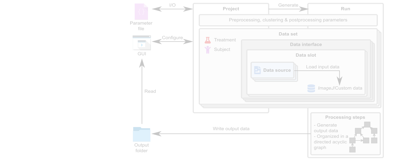
Our MSOT cluster analysis toolkit enables the quantitative and automated analysis of multispectral optoacoustic tomography (MSOT) images. The pharmacokinetics of…


