

Deaths due to liver disease have increased significantly. The liver is made up of key structures that functional components of the liver. Sinusoids are specialised porous blood vessels found mainly in the liver. They are a type of capillary that differs from typical capillaries because of their larger diameter and more permeable walls. The unique structure of sinusoids allows efficient exchange of substances between the blood and surrounding tissue. Kupffer cells are liver macrophages that line the sinusoids and play a crucial role in filtering pathogens and debris from the blood. Canaliculi are microscopic channels of particular importance in the liver, where they play a crucial role in bile flow and detoxification. Hepatocytes are the primary functional cells of the liver, crucial for metabolic processes such as gluconeogenesis, lipid metabolism and detoxification, as well as synthesising plasma proteins and producing bile for digestion. They also play an important role in vitamin storage and the detoxification of ammonia into urea for excretion. Polyplex micelles are essential in liver treatment for delivering therapeutic agents like siRNA.Their design allows targeted delivery to hepatocytes for the treatment of genetic defects, viral infections and liver pathologies.
Therefore, for research hepatocytes are the primary target because of their central role in liver function and disease. Canaliculi and sinusoids are critical for understanding how micelles traverse the intricate architecture of the liver, while Kupffer cells are being studied for their role in nanoparticle clearance and immune response modulation. These analyses are critical for optimising micelle design to enhance hepatocyte-specific delivery while evading immune clearance, thereby improving therapeutic outcomes in liver disease.
In this study, hepatocytes, canaliculi, sinusoids and Kupffer cells are analysed, to understand the difference in uptake of various polyplex micelles.
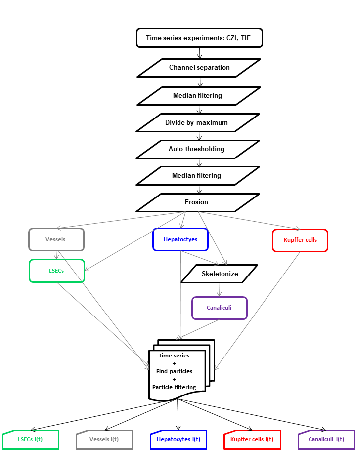
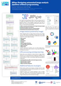
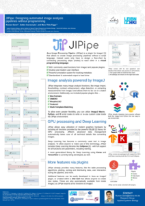
The previously established automated image analysis algorithm adapted to the situation at hand, calculated the retention profile of each polyplex micelle in the sinusoids. The automated image analysis process described uses JIPipe based on ImageJ 1.53q to analyse confocal images of liver tissue, focusing on hepatocytes, canaliculi, sinusoids by autofluorescence and Kupffer cells by staining. Pre-processing includes noise removal, normalisation, auto thresholding, despeckling and specific erosion steps to outline hepatocytes, canaliculi and sinusoids. Segmentation starts with autofluorescence images for hepatocytes and canaliculi, and refines the areas by subtracting canaliculi regions (as provided by a skeletonisation algorithm). Sinusoids are segmented similarly, but with darker area thresholds and more erosion (based on the average projection of the entire time series). Kupffer cells are segmented based on their staining characteristics and proximity to sinusoids.
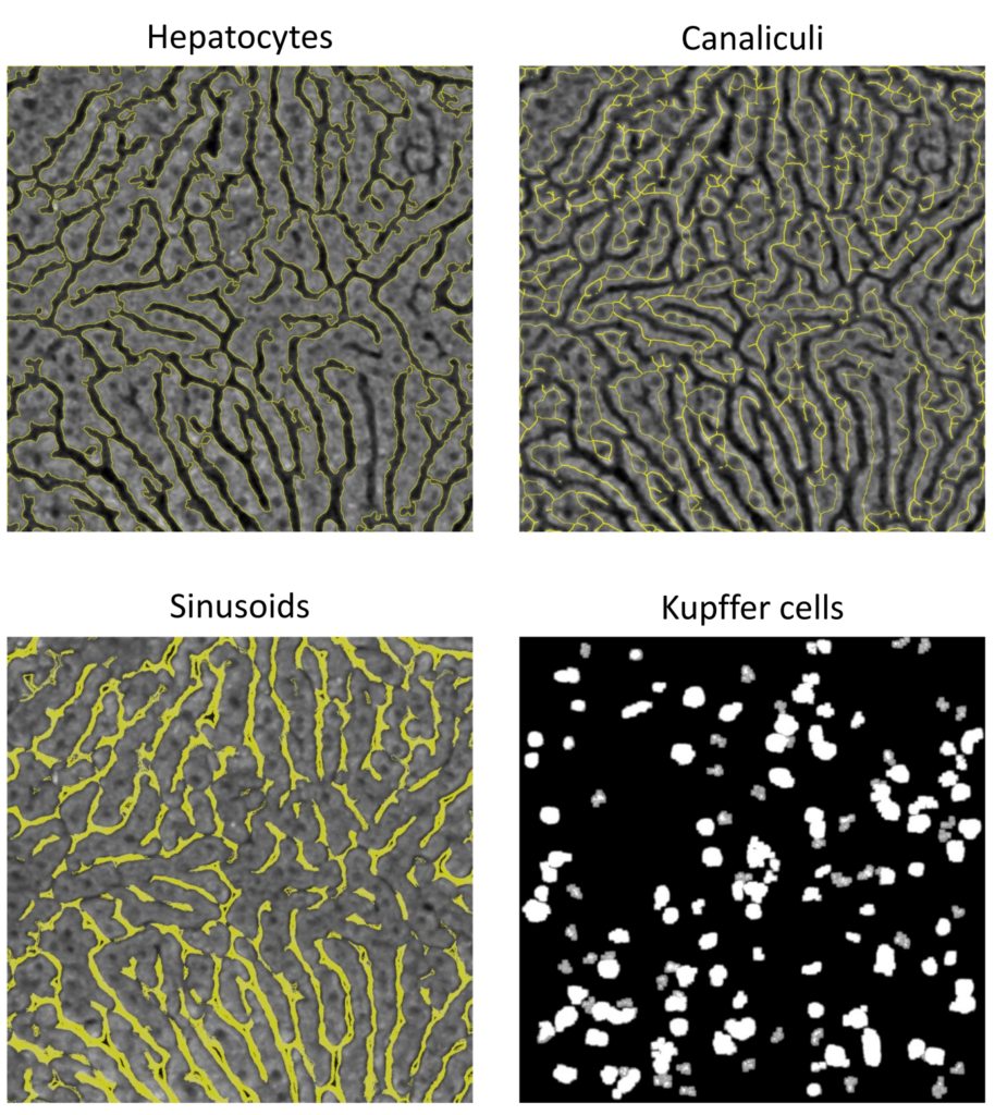
Image analysis revealed different patterns of accumulation of different polyplex micelles in hepatocytes, ranging from effective targeting to broader distribution throughout hepatocytes and non-parenchymal cells, suggesting potential interactions with multiple cell types within the liver.
Experiemntal Collaborators
Department of Anesthesiology and Intensive Care Medicine at the University Hospital, Jena
Jena Center for Soft Matter (JCSM) at the Friedrich Schiller University in Jena
Leibniz Centre for Photonics in Infection Research at the Leibniz Institute of Photonic Technology in Jena
Center for Sepsis Control and Care at the University Hospital, Jena