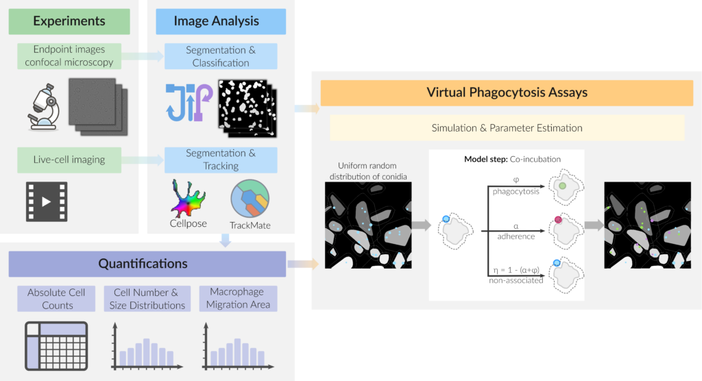
Confrontation and phagocytosis assays are commonly used to investigate host-pathogen interactions of certain immune cells, such as phagocytes like macrophages or neutrophils and various pathogens. Traditional phagocytosis assays, which make use of techniques such as imaging flow cytometry, confocal microscopy, and live cell imaging, have limitations due to their population-based nature. These traditional measures, including the phagocytosis ratio and uptake ratio, fail to provide microscopic parameters and can give ambiguous results. To address these issues, we developed a novel approach combining automated image analysis and individual-based modeling to simulate virtual phagocytosis assays based on a modeling framework, CellRain, which performs Monte-Carlo simulations on endpoint images and therefore is able to provide such microscopic parameters.
Virtual Phagocytosis Assay use automated image analysis combined with individual-based modeling to simulate and characterize the phagocytosis process.

The virtual phagocytosis assay is a computer-based simulation that characterizes the engulfment of pathogens by macrophages, based on experimental images. The images were analysed to determine the dimensions of the cells and spores and their distributions. The simulation randomly distributes virtual spores around the macrophages, using the aforementioned images as a basis. The model simulates the probabilities of phagocytosis represent the interactions between macrophages and spores. The analysis revealed strain-specific differences in phagocytosis and macrophage migration, thereby highlighting limitations and ambiguities in traditional population-based phagocytosis measures.
Experimental Collaborators
- Infection Biology and Molecular Biotechnology at the at the Leibniz-HKI in Jena, Germany
- Microbiological Research Network of Friedrich-Schiller-University Jena, Germany
- Institute for Organic Chemistry and Macromolecular Chemistry of Friedrich-Schiller-University Jena, Germany


