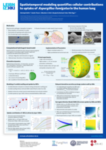
The first line of defense against the airborne human-pathogenic fungus A. fumigatus are the resident immune cells in the human lung – the alveolar macrophages. A. fumigatus conidia have to be cleared from the lung before onset of germination, which occurs approximately six hours after entry in the lung. Thus, understanding the host-pathogen interaction between A. fumigatus and macrophages is an important step along the road towards the development of new diagnosis and treatment strategies against this fungal pathogen. Modeling can be of great help in this regard, since parameters can be predicted that are not directly accessible from experiment. Different techniques can be applied such as agent-based modeling, evolutionary game theory or performing Monte-Carlo simulations on microscopic images.
Experiments
Confrontation assays with alveolar macrophages and different A. fumigatus strains have been performed. Conidia were differentially stained before and after 1 hour of co-incubation to distinguish phagocytosed from non-phagocytosed conidia. After 1 hour of co-incubation fluorescence microscopy images were taken (endpoint images). Furthermore, live cell microscopy was performed for confrontation assays with macrophages and the different A. fumigatus strains.
Experimental Collaborators
- Molecular and Applied Microbiology at the at the Leibniz-HKI in Jena, Germany
- Jena Microbial Resource Collection at the at the Leibniz-HKI in Jena, Germany
Image analysis
Image analysis of endpoint images involved segmentation and classification of macrophages and conida. This was performed using ACAQ. Live cell image data was used to segment and track macrophages in order to calculate the area macrophages are scanning in a certain time interval. For cell segmentation Cellpose was used and tracking was done using AMIT.
Model
Modeling was performed using the framework CellRain on the endpoint images with segmented macrophages. Furthermore, quantitative measures derived from the image analysis was used to calibrate the model and to estimate model parameters. These microscopic parameters can be compared for various strains.
