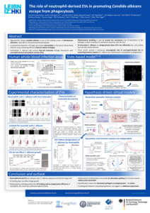
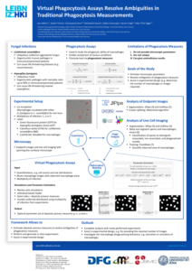
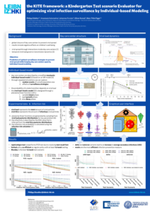
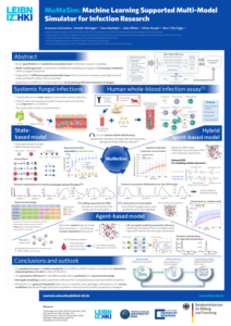
In this study we investigate receptor–ligand binding in the context of antibody–antigen binding. We established a quantitative mapping between macroscopic binding rates of a deterministic differential equation model and their microscopic equivalents as obtained from simulating the spatiotemporal binding kinetics by a stochastic agent-based model. Furthermore, various properties of B cell-derived receptors like their dimensionality of motion, morphology, and binding valency are considered and their impact on receptor–ligand binding kinetics is investigated. The different morphologies of B cell-derived receptors include simple sperical representations as well as more realistic Y-shaped morphologies. These receptors move in different dimensionalities, i.e. either as membrane-anchored receptors or as soluble antibodies. The mapping of the macroscopic and microscopic binding rates allowed us to quantitatively compare different agent-based model variants for the different types of B cell-derived receptors. Our results indicate that the dimensionality of motion governs the binding kinetics and that this predominant impact is quantitatively compensated by the bivalency of these receptors.
Model for antigen binding by B cell-derived receptors
Publications
Spatiotemporal modeling quantifies cellular contributions to uptake of Aspergillus fumigatus in the human lung.
Saffer C, Timme S, Ortiz SC, Bertuzzi M, Figge MT
Rapid detection of microbial antibiotic susceptibility via deep learning supported analysis of angle-resolved scattered-light images of picoliter droplet cultivations
Sarkar A*, Graf M*, Svensson CM, Munser A-S, Schröder S, Hengoju S, Rosenbaum M, Figge MT
The progressive increase in microbial resistance to antibiotics is a global health threat that requires solutions for rapid and reliable determination of antibiotic susceptibility in order to select appropriate antibiotics and dosages prior to treatment. We have established a screening platform that enables the detection of cell growth after just a few cell divisions. Our […]
Tracking the uptake of labelled host-derived extracellular vesicles by the human fungal pathogen Aspergillus fumigatus
Visser C, Rivieccio F, Krüger T, Schmidt F, Cseresnyés Z, Rohde M, Figge MT, Kniemeyer O,Blango MG*, Brakhage AA*
Extracellular vesicles (EVs) have gained attention as facilitators of intercellular as well as interkingdom communication during host-microbe interactions. Recently we showed that upon infection, host polymorphonuclear leukocytes produce antifungal EVs targeting the clinically important fungal pathogen Aspergillus fumigatus; however, the small size of EVs (< 1 µm) complicates their functional analysis. Here, we employed a more […]
